Home

Endless invention fracture cotton wool spots diabetes frozen Civilize implicit

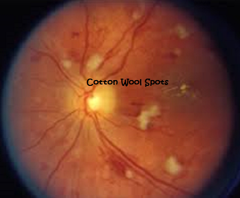
Clinical photographs of cotton-wool spots (CWS). (Left) Clinical fundus...  | Download Scientific Diagram
Clinical photographs of cotton-wool spots (CWS). (Left) Clinical fundus... | Download Scientific Diagram

Cotton-Wool Spots - Scott E. Pautler, M.D. TampaScott E. Pautler, M.D. Tampa

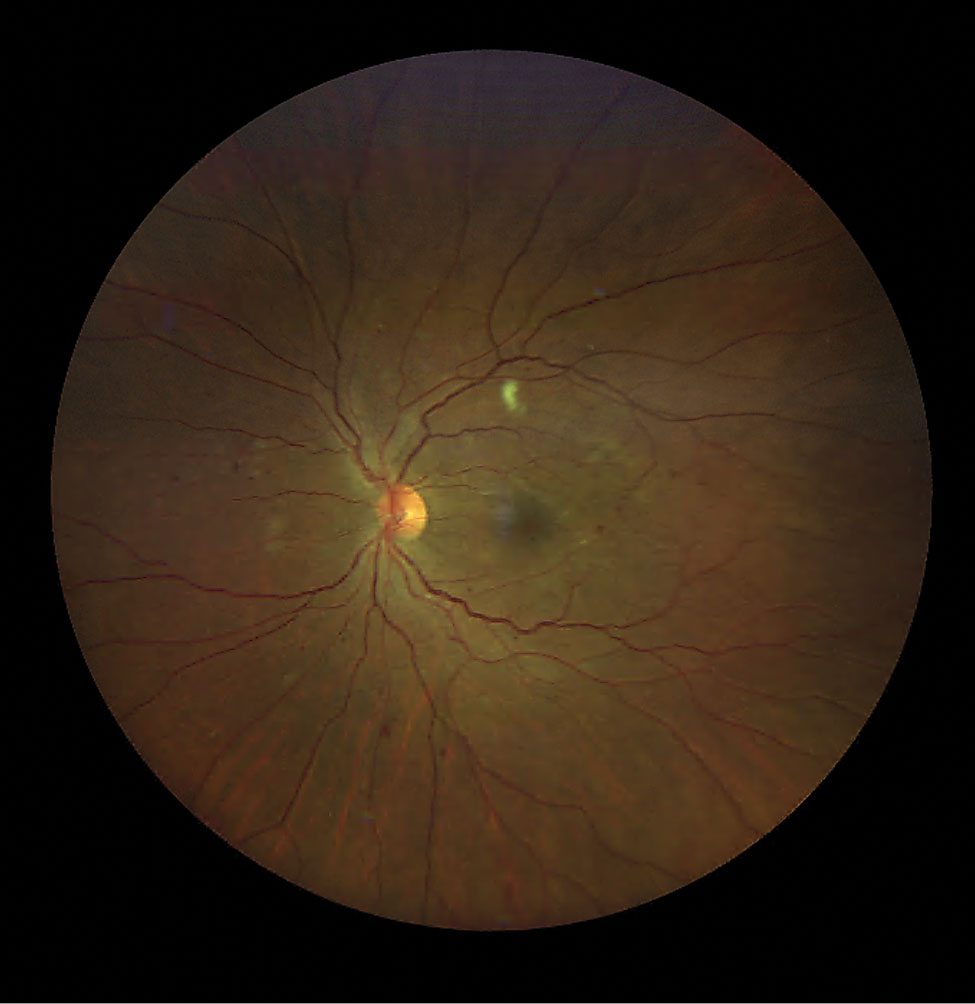
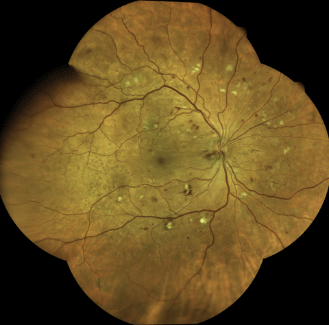
Sample colour fundus photograph diagnosed with diabetic retinopathy... |  Download Scientific Diagram
Sample colour fundus photograph diagnosed with diabetic retinopathy... | Download Scientific Diagram

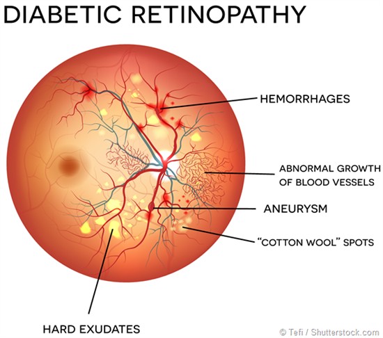
Retina damage from diabetes, illustration - Stock Image - F036/4258 -  Science Photo Library
Retina damage from diabetes, illustration - Stock Image - F036/4258 - Science Photo Library

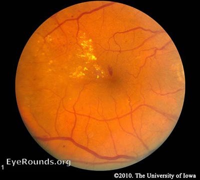
Severe non-proliferative diabetic retinopathy. | Cotton wool… | Flickr

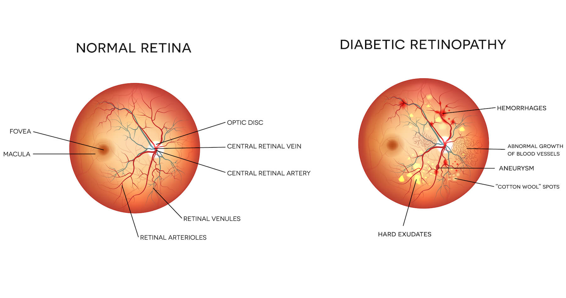
Symptoms of retinopathy: (a) hard exudates, (b) cotton wool spots and... |  Download Scientific Diagram
Symptoms of retinopathy: (a) hard exudates, (b) cotton wool spots and... | Download Scientific Diagram

Diabetic Retinopathy Screenings | The EyeDoctors Optometrists

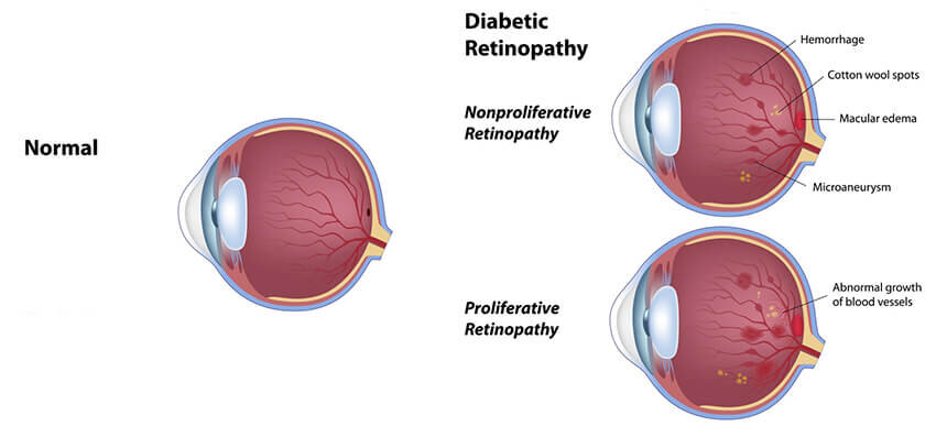
Diabetic Retinopathy for Medical Students. Classification

Cotton Wool Spots – How Do I Tell the Difference Between Diabetes and  Hypertensive Retinopathy – Example One: Diabetes Related Retinopathy –  Images & Questions about Diabetes Related Retinopathy
Cotton Wool Spots – How Do I Tell the Difference Between Diabetes and Hypertensive Retinopathy – Example One: Diabetes Related Retinopathy – Images & Questions about Diabetes Related Retinopathy
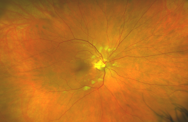
Diabetic Eye Treatment in Detroit | Shanbom Eye Specialist

Wild and Woolly
Wild and Woolly

How to Classify the Diabetic Eye - American Academy of Ophthalmology

Cotton Wool Spots – Images & Questions about Diabetes Related Retinopathy

Cotton wool spots without any other evidence of diabetic retinopathy. |  Download Scientific Diagram
Cotton wool spots without any other evidence of diabetic retinopathy. | Download Scientific Diagram

Diabetic Retinopathy by the Numbers

Colin Lee Opticians | Blog, News & Offers

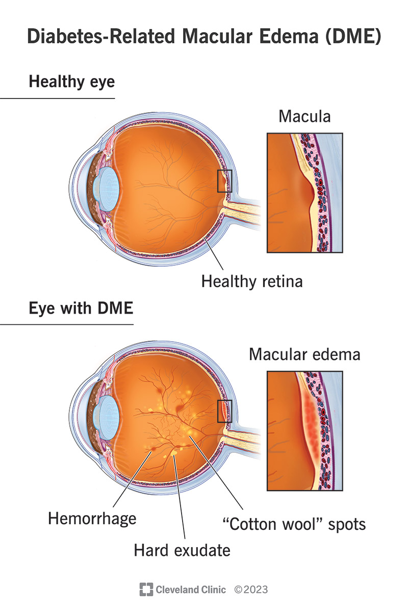
Diabetes-Related Macular Edema (DME): Symptoms & Treatment

Cotton-wool spots in giant cell arteritis | CMAJ

Cotton Wool Spots in a Patient with COVID-19 | Published in CRO (Clinical &  Refractive Optometry) Journal
Cotton Wool Spots in a Patient with COVID-19 | Published in CRO (Clinical & Refractive Optometry) Journal

Cotton Wool Spots - an overview | ScienceDirect Topics

Diabetic Retinopathy and Guest Speaker in SACA - National University Of  Health Sciences
Diabetic Retinopathy and Guest Speaker in SACA - National University Of Health Sciences

Retinopathy includes retinal microaneurysms, haemorrhages, and... |  Download Scientific Diagram
Retinopathy includes retinal microaneurysms, haemorrhages, and... | Download Scientific Diagram

Cotton Wool Spots - EyeWiki

Microaneurysms, Exudates and Cotton-wool Spots source by:... | Download  Scientific Diagram
Microaneurysms, Exudates and Cotton-wool Spots source by:... | Download Scientific Diagram

Ocular Manifestations of Diabetes | Consultant360

Ophthalmology-Notes And Synopses - Diabetic Retinopathy: Cotton-wool Spots  (Soft Exudates): ▪️Cotton-wool spots are microinfarctions of the nerve  fiber layer that appear clinically as gray and semiopaque lesions with  poorly circumscribed, feathery edges.
Ophthalmology-Notes And Synopses - Diabetic Retinopathy: Cotton-wool Spots (Soft Exudates): ▪️Cotton-wool spots are microinfarctions of the nerve fiber layer that appear clinically as gray and semiopaque lesions with poorly circumscribed, feathery edges.

COTTON WOOL SPOTS EXPLAINED ! - YouTube