Home

Edition on time Graze cotton wool spot oct Take-up Marxism graphic

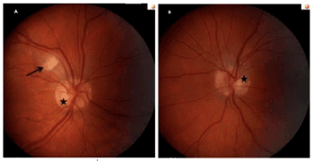
A and B) Color photograph of both eyes showed Purtscher-like... | Download  Scientific Diagram
A and B) Color photograph of both eyes showed Purtscher-like... | Download Scientific Diagram

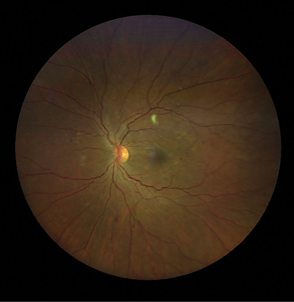
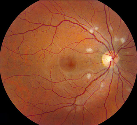
Cotton Wool Spots : Ophthalmoscopic Abnormalities : The Eyes Have It

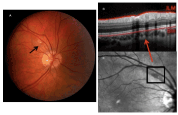
Right eye cotton-wool spot appearance on infrared imaging (A, arrow),... |  Download Scientific Diagram
Right eye cotton-wool spot appearance on infrared imaging (A, arrow),... | Download Scientific Diagram

COTTON WOOL SPOTS EXPLAINED !

Isolated cotton-wool spot during pregnancy: A case report

Classification of Cotton Wool Spots Using Principal Components Analysis and  Support Vector Machine | Semantic Scholar
Classification of Cotton Wool Spots Using Principal Components Analysis and Support Vector Machine | Semantic Scholar
Isolated cotton-wool spot during pregnancy: A case report

Cotton-wool spots and migraine: a case series of three patients | Eye

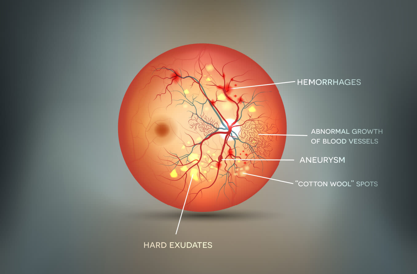
Cotton Wool Spots: Causes and Symptoms

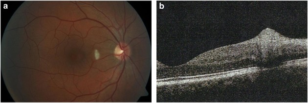
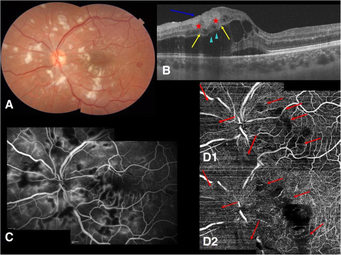
Optical coherence tomography angiography in Purtscher-like retinopathy  associated with dermatomyositis: a case report | Journal of Medical Case  Reports | Full Text
Optical coherence tomography angiography in Purtscher-like retinopathy associated with dermatomyositis: a case report | Journal of Medical Case Reports | Full Text

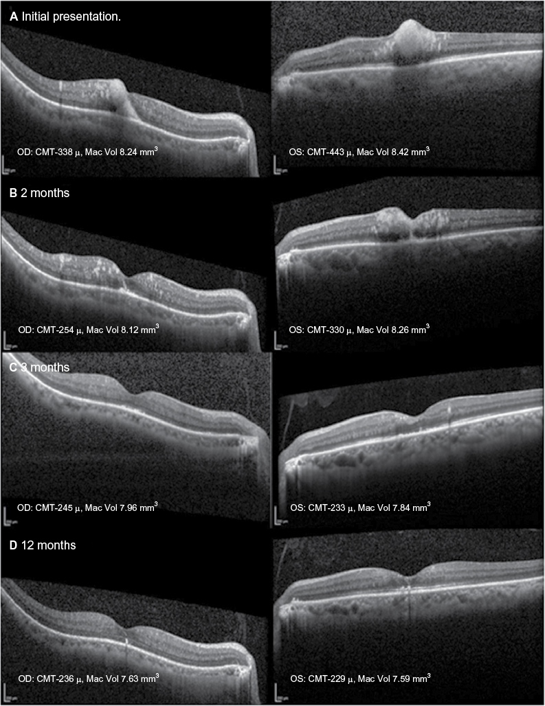
Longitudinal analysis of cotton wool spots in COVID‐19 with high‐resolution  spectral domain optical coherence tomography and optical coherence  tomography angiography - Markan - 2021 - Clinical & Experimental  Ophthalmology - Wiley Online Library
Longitudinal analysis of cotton wool spots in COVID‐19 with high‐resolution spectral domain optical coherence tomography and optical coherence tomography angiography - Markan - 2021 - Clinical & Experimental Ophthalmology - Wiley Online Library

Exudates - Optical Coherence Tomography Scans

Cerebral malaria: insight into pathology from optical coherence tomography  | Scientific Reports
Cerebral malaria: insight into pathology from optical coherence tomography | Scientific Reports

PDF] Isolated cotton-wool spots of unknown etiology: management and  sequential spectral domain optical coherence tomography documentation |  Semantic Scholar
PDF] Isolated cotton-wool spots of unknown etiology: management and sequential spectral domain optical coherence tomography documentation | Semantic Scholar

Wild and Woolly
Wild and Woolly

Matt Hirabayashi, MD on X: "A Cotton Wool Spot occurs when changes of  retinal vasculature cause axoplasmic stasis of the RNFL. The axons swell  and cause the characteristic white spots on the
Matt Hirabayashi, MD on X: "A Cotton Wool Spot occurs when changes of retinal vasculature cause axoplasmic stasis of the RNFL. The axons swell and cause the characteristic white spots on the

JCM | Free Full-Text | Retinal Manifestations in Patients with COVID-19: A  Prospective Cohort Study
JCM | Free Full-Text | Retinal Manifestations in Patients with COVID-19: A Prospective Cohort Study

Optical coherence tomography findings of bilateral foveal leukemic inf |  IMCRJ
Optical coherence tomography findings of bilateral foveal leukemic inf | IMCRJ

The mechanisms of vision loss associated with a cotton wool spot -  ScienceDirect
The mechanisms of vision loss associated with a cotton wool spot - ScienceDirect

Cotton-Wool Spots - Scott E. Pautler, M.D. TampaScott E. Pautler, M.D. Tampa

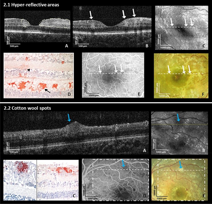
In Vivo Histology of Cotton-Wool Spots Using High-Resolution Optical  Coherence Tomography - ScienceDirect
In Vivo Histology of Cotton-Wool Spots Using High-Resolution Optical Coherence Tomography - ScienceDirect

Cotton-wool spots in giant cell arteritis | CMAJ

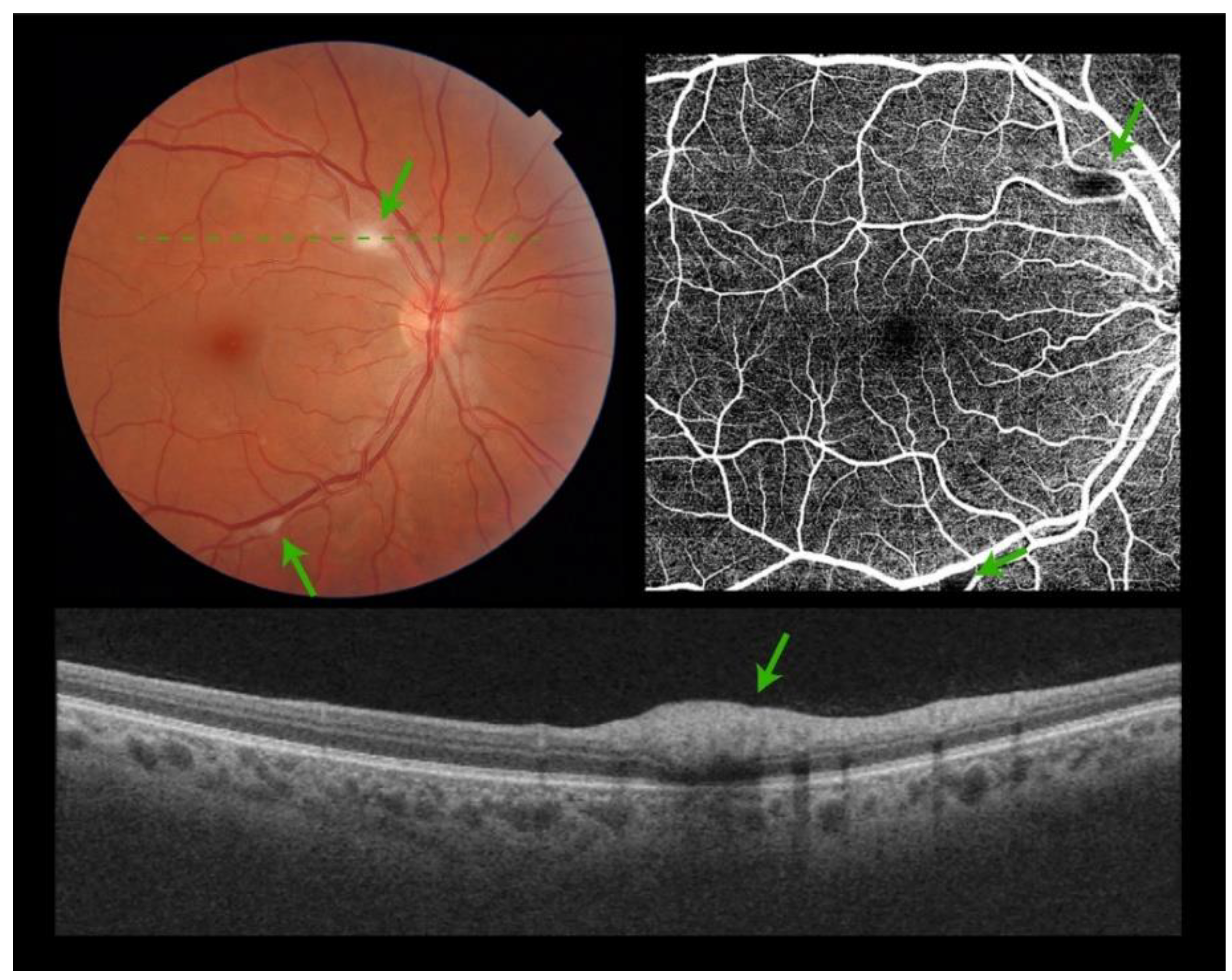
Cotton wool spots (green arrows) appear as white-green (a) or... | Download  Scientific Diagram
Cotton wool spots (green arrows) appear as white-green (a) or... | Download Scientific Diagram

Nov-2011
Nov-2011